Abstract
Provided here methods of generating human alveolar macrophage-like cells in-vitro from blood-derived monocytes by culturing them in a cell culture medium containing a mixture of a surfactant and two or more cytokines. This mixture can contain calfactant, granulocyte-macrophage colony-stimulating factor, transforming growth factor beta, and interleukin-10.
Application Number
17657344 (non-provisional)
Date of Application
March 30, 2022
Inventors
Larry S. Schlesinger and Susanta Pahari
In Vitro Differentiation of Human Alveolar Macrophages from Blood Monocytes
Description
Human alveolar macrophages (a type of white blood cell) are the first line of defense against invading respiratory pathogens. Human alveolar macrophages are critical for tissue homeostasis, host defense, clearance of surfactant and cell debris, pathogen recognition, initiating and resolving lung inflammation, and repairing damaged tissue. However, in respiratory diseases (collectively valued at 35.6 billion in 2017), including
chronic obstructive pulmonary disease (COPD), asthma, cystic fibrosis, tuberculosis, and COVID-19, human alveolar macrophages appear to be dysfunctional and may contribute to disease pathogenesis. However, research into this phenomenon has been complicated and hindered by the fact that these cells are only available from human donors by bronchoalveolar lavage or from recently deceased individuals, and the cell phenotype rapidly changes during ex vivo tissue culture conditions.
Inventors at Texas Biomed have developed a unique culture condition that allows for the conversion of blood-obtained monocytes to a human alveolar macrophage phenotype. By identifying and mimicking components of the alveolar microenvironment, the inventors have created a unique cocktail of three cytokines and a surfactant that differentiates and preserves Alveolar Macrophage Like (AML) cells that retain human alveolar macrophage function in tissue culture.
Benefits and Advantages:
- Provides a readily accessible in vitro AML model to study the impact of human alveolar macrophages on infectious and inflammatory processes and diseases, as well as testing potential therapeutics for drug discovery.
- Cheaper and less labor intensive than current methods to obtain human alveolar macrophages from bronchoalveolar lavage or cadavers.
- The protocol to differentiate blood-obtained monocytes to AML cells is simple, can be completed in 6 days, and could easily be packaged as a kit.
- Generated AML cells show a high degree of similarity to human alveolar macrophages obtained by conventional means as characterized by gene expression, marker staining and functional studies.
- Unlike alveolar macrophages removed from lungs, generated AML cells retain their phenotype in vitro, making longer-term and mechanistic studies a possibility for researchers.
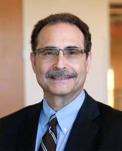
LARRY S. SCHLESINGER, M.D.
President/CEO, Professor
Dr. Schlesinger is an internationally recognized authority in infectious diseases with a particular interest in tuberculosis and lung biology. As a physician scientist, his studies focus on the pathogenesis of tuberculosis and other airborne infectious agents that subvert lung immune mechanisms. Tuberculosis is the deadliest infectious disease in the world.

Inside The Lab
Dr. Schlesinger and his research team have made major discoveries about the human immune response to pathogens, and he is translating these into host-directed drug discovery platforms. His laboratory studies innate immunity to these infectious pathogens by focusing on their interactions with human mononuclear phagocytes, which are cells that protect the body by ingesting harmful cells, bacteria or foreign particles.
Another focus of Dr. Schlesinger’s work is understanding how the alveolar (air compartments of the lung) environment affects the biology of alveolar macrophages (a type of white blood cells) in ways that directly impact the host response to airborne infectious agents.
Dr. Schlesinger is also interested in the impact of diabetes and aging on the immune response to M. tuberculosis, susceptibility to infection, and new imaging and drug discovery platforms for mycobacteria.
As a physician scientist, I pursue research directions that enable me to discover something that translates into helping to improve human lives. Texas Biomed is a friendly, collaborative environment with outstanding research and limited bureaucracy for getting things accomplished. My passionate is in building great research enterprises where the next generation can learn and
grow.
—Dr. Larry Schlesinger
Texas Biomedical Research Institute
The institute pioneers and shares scientific breakthroughs that protect you, your families and our global community from the threat of infectious diseases. Texas Biomed is an independent, not-for-profit, research institute with a strong history of collaborating with global partners and contributing to the world of science and human health for more than 80 years. We are evolving into a one-of-a-kind, worldleader in infectious diseases, with researchers working together in three critical areas of discovery. Our partnerships continue to expand globally to deliver new diagnostics, treatments and cures through our preclinical research and scientific
programs.
Scientific Programs
Host-Pathogen Interactions (HPI) – The HPI Program concentrates on the basic biology of infection in humans and animals and the development of disease.
Disease Intervention & Prevention (DIP) – The DIP Program aids in the development of diagnostics, treatments and vaccines to prevent disease, reduce severity of disease and infection, if not cure infection.
Population Health (PH) – The PH program aims to identify correlates of disease susceptibility or resistance to infectious diseases on a population level
Resource Attributes
- BSL-3 and BSL-4 facilities with laboratory capacity to perform in vitro experiments (biochemistry, cell biology, molecular genetics, and immunology) and in vivo studies (vaccine/therapeutic efficacy, pathogenesis and survival) including imaging.
- Experience with the development of nonhuman primate (baboon, macaque, marmoset) and rodent (mouse, hamster, and Guinea pig) models for evaluating disease pathology and diagnostic, therapeutic or vaccine efficacy.
- Established standard operating procedures and protocols for biochemical, immunological and metabolism assays, deep sequencing, neutralization assays, determining bacterial and viral load among others.
- Facilities equipped with state-of-the-art equipment including Aerobiology instrumentation, Telemetry, Clinical Pathology Instrumentation, Flow Cytometry, Sorting, PET-CT Scan, Live-Imaging, and Single-Cell Sequencing among others.
- Active programs with NIH/DOD/BARDA, commercial and international partners.
- Studies adhering to robust quality standards to support regulatory filings with the FDA and other agencies.
For more information on Texas Biomed, visit www.txbiomed.org. To learn more about partnering with our world-class researchers to execute quality scientific studies, please contact Applied Science and Innovation at